
Analytes: 4 of 6
Extracellular Vesicles And Exosomes
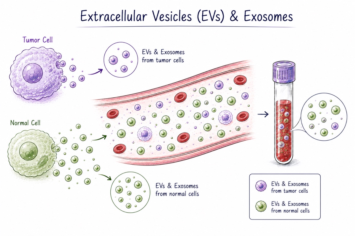
Cells do more than leak material when they die. They also package material and release it on purpose. These packages are tiny bubbles surrounded by a membrane similar to the membrane around the cell itself. They are called extracellular vesicles. Exosomes are one well-studied type of extracellular vesicle.
An extracellular vesicle can carry proteins, RNA, sometimes DNA, and fats from the cell that made it. Cells use these packages to communicate with other cells. Tumors appear to use them too, including to influence tissue in distant parts of the body before cancer spreads there.
For liquid biopsy, the wrapping is useful. It protects material that might otherwise be destroyed in the bloodstream. The outside of the vesicle also carries markers from the parent cell, so in principle researchers may be able to sort vesicles by where they came from and then examine the ones from a particular tissue. That could help separate a disease signal from the much larger amount of material released by the rest of the body.
Actually separating the vesicles is difficult. Blood plasma is full of other particles that are almost the same size, especially particles that carry fats around the body, and there are far more of those. Laboratories also use different methods to isolate vesicles, which means two studies describing "exosomes" may not be examining exactly the same mixture. Even the names for some vesicle subgroups are still debated. All of that makes studies harder to compare.
In this index: extracellular vesicles appear in 6,351 of the classified papers and patents.
This page explains what these tests are and how researchers use them. It is not medical advice, and it is not a guide to whether any test is right for you or for a patient. Talk to a clinician about testing decisions.